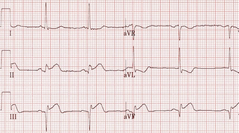

The authors conclude that although the negative predictive value may be at least modest, the presence of Q or QS complexes on ECG should not be used to determine the presence of myocardial scar or to guide in whom coronary revascularization should or shouldn’t be sought 8. On a per vessel analysis these were 56%, 73%, 25%, and 91%, respectively, for Q waves and 33%, 87%, 30%, and 89%, respectively, for only QS complexes.

When using only QS complexes, the corresponding vales were 46%, 59%, 36%, and 68% respectively. On a per patient analysis, the sensitivity, specificity, positive predictive value (PPV), and negative predictive value (NPV) of Q waves for detecting scar were 70%, 40%, 37%, and 73% respectively. The presence of Q waves was associated with a higher number of scarred segments on PET (2.0 vs 1.7). They evaluated the correlation of Q and/or QS waves on ECG with scarred myocardium by PET defined as a region of matching defect (<50% uptake) on FDG and ammonia PET images. They studied 149 patients known to have ischemic cardiomyopathy defined by evidence of coronary artery stenosis on prior angiography or a history of MI who underwent ammonia and FDG PET at a single institution. In this issue of the Journal, Markendorf and colleagues evaluated the ability of ECG to predict the presence of myocardial scar using cardiac PET as gold standard 8. Other conditions that have been associated with Q waves in the absence of MI include Wolff-Parkinson-White syndrome, amyloid heart disease, hypertrophic cardiomyopathy, non-ischemic dilated cardiomyopathy, chronic obstructive pulmonary disease, left bundle branch block, and paced rhythm 2. Any condition that induces a shift in the position of the heart in relation to the surface ECG leads can have similar consequences. Notoriously, lead misplacement can result in the misdiagnosis of MI by ECG. Even pathologic Q waves may be present in the absence of MI. In this regards, not all Q waves on ECG tracings are pathologic pathologic Q waves are defined in the Fourth Universal Definition of Myocardial Infarction as the presence of Q wave in leads V2-V3 > 0.02 s or QS complex in leads V2-V3 Q wave ≥ 0.03 s and ≥ 1 mm deep or QS complex in leads I, II, aVL, aVF, or V4-V6 in any 2 leads of a contiguous lead grouping (I, aVL V3-V6 II, III, aVF) or an R wave > 0.04 s in V1-V2 and R/S > 1 with a concordant positive T wave in absence of conduction defect 3. Nevertheless, the presence of Q waves on an ECG is helpful to suggest the presence of prior MI. Further, we now realize that transmural MI may occur without Q waves on the ECG and non-transmural MIs may be accompanied by Q waves 2. Although prior guidelines used to classify MI as Q-wave and non-Q-wave MIs, current guidelines rely on ST segment shift on the ECG to differentiate ST-elevation MI from non-ST elevation acute coronary syndrome since that has direct impact on clinical management 1. Surface electrocardiography (ECG) is often used clinically for the detection of myocardial infarction (MI).


 0 kommentar(er)
